- Review
- Open access
- Published:
Microsponges: a breakthrough tool in pharmaceutical research
Future Journal of Pharmaceutical Sciences volume 8, Article number: 31 (2022)
Abstract
Background
A microsponge delivery system (MDS) is an innovative and unique way of delivering drugs in a structured manner. Using microsponge drug delivery, regulated drug delivery may now be achieved quickly and easily.
Main body
MDS comprises porous microspheres ranging in size from 5 to 300 microns, with a large porous structure and a very tiny spherical shape. MDS is normally used to deliver drugs via topical channels, but they have recently shown the potential approach to drug delivery via oral, ophthalmic and parenteral routes. MDS can easily modify the pharmaceutical release contour and improve formulation stability while minimising the negative impact of the drug. The fundamental purpose of microsponge drug administration is to reach the highest possible peak plasma concentration in the blood. The capacity of MDS to self-sterilise is their most prominent attribute.
Conclusions
MDS is used as anti-allergic, anti-mutagenic and non-irritant in innumerable investigations. This review includes formulation, criteria for drugs to be incorporated in MDS, formulation methods, assessment parameters, and role of MDS in the management of various disorders. This review will be quite useful in the future in exploring the MDS in different disorders.
Graphical Abstract

Background
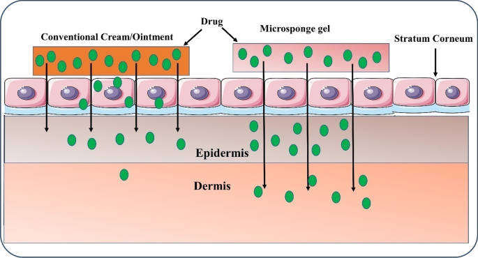
Drug release techniques are designed particularly for the purpose of delivering drugs to specific places inside the body. This increases the efficiency of drug treatment, and patient compliance has a substantial influence on the health care system [1]. The pharmaceutical industry has learned that delivering APIs to controlled target locations within the body has proven to be a significant issue [2]. Recently, much attention has been dedicated to the creation of novel drug delivery systems based on microsponges, as well as the modification and regulation of drug release performance. By combining a drug with a carrier system, it may be feasible to vary the duration and therapeutic index of the drug [3]. The most consistent and predictable route for active substances to enter the body's local and systemic systems is through the skin, as shown in Fig. 1. The major drawback of the transdermal administration route is that a common route of pharmaceuticals is poorly soluble in water, causing various obstacles during the creation of conventional dosage forms. Another important difficulty with topical pharmaceutical administration is evaporation of the drug, unpleasant fragrance, and visually unattractive carriers, which can result in greasiness and stickiness and may lead to impaired patient compliance [3]. Due to their ineffectiveness, traditional methods demand a larger amount of drug be incorporated for beneficial therapy. MDS based on polymeric microspheres may be able to achieve this objective [4]. Won created the MDS drug delivery method in 1987; his patents were assigned to Advance Polymer Systems, Inc. Following that, Cardinal Health, Inc. was awarded a licence to use this innovative technology in topical treatments. This corporation launched a huge number of improvements in approach and applied them to over-the-counter items, cosmetics, and pharmaceutically prescribed products [4]. Traditional topical medicines are meant to have an influence on the outer layer of the skin. Following the application of such mixtures, their active moieties are released. They provide a film of concentrated active moiety, which is readily absorbed. This leads to an increased accumulation of medicines in the dermis. MDS can assist to avoid this. Perhaps, the microsponge drug delivery technology can considerably limit drug unwanted effects like itching while yet delivering the medicine efficiently to the skin [5]. Due to their visually unpleasant appearance, as well as their stickiness and greasiness, patients' adherence to ointments is low. On the other hand, the MDS prolongs the time, which drug spends on the surface of the skin. Microsponge delivery technology outperforms other strategies such as microencapsulation and liposomes in terms of quality. Liposomes have a limited carrying capacity, are difficult to make, and are chemically and microbially fragile. The rate of release of the pharmaceutical active moiety is routinely regulated in microencapsulation. When the microcapsule wall is ruptured, the pharmaceutically active moiety is released [5]. MDS porous polymeric microspheres are used to extend the life of topical drugs. It is claimed that they release the drug at the smallest practical dose in the most effective manner possible, resulting in greater stability and the capacity to reduce undesirable effects and change the drug release profile. MDS is capable of absorbing secretions, which results in less greasy skin. These are microscopic, inert, and unbreakable spheres that cannot penetrate the skin. They collect to a certain extent in the skin's minute crevices and nooks and release the drug contained within the MDS when the skin needs it. MDS' microspheres size (diameter of 5–300 µm) may vary according to the smoothness of the skin [6, 7]. MDS can assist in avoiding an excessive build-up of components in the skin [7]. Numerous perfumes, essential oils, sunscreens, antifungal, emollient, and anti-infective agents, as well as pharmaceuticals, become trapped in MDS. MDS is stable at pH values ranging from 1 to 11 and temperatures up to 130 °C. The vast majority of vehicles and substances are compatible with the components of MDS. Bacteria cannot penetrate a 0.25-µm-diameter MDS pore, which makes MDS compositions self-sterilising [4, 8].
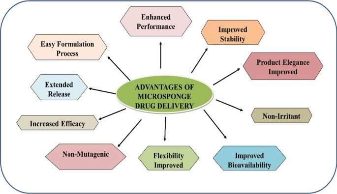
The drug must be totally mixable in the monomer or can be made mixable by adding a small quantity of non-polar solvent. It must be insoluble in water and inert to monomers. Otherwise, the MDS will deteriorate prior to application, and the vehicle must be stable in proximity to and under specified circumstances of the polymerization catalyst [4, 9]. As shown in Fig. 2, MDS technology possesses various advantages over other drug delivery systems [4, 10, 11].
Main text
A number of polymers were utilised to form the MDS ‘cage.’ Eudragit RS-100, polylactide-co-glycolic acid, Eudragit RS PO, polydivinylbenzene, Eudragit S-10, polylactic acid, and polyhydroxybutyrate are among the polymers explored for the creation of MDS for utilisation topically or orally. The polymer Eudragit RS-100 was the most commonly examined polymer due to its versatility in allowing the investigator to apply it in a number of ways. By improving the solubility, the polymer Eudragit RS PO also controls drug release. Polymers such as polylactic acid and polylactide-co-glycolic acid were also studied for peptide and protein delivery. MDS generated with these polymers also exhibits the ability to float due to the hydrophobicity of the polymer, by preventing the particles from becoming wet in aquatic circumstances, allowing them to be utilised to make floating MDS. The usage of polymers in the fabrication of MDS reveals that production techniques may be modified to satisfy unique demands. A few MDS formulations further incorporated a plasticizer, such as triethyl citrate, for the active moiety and polymers, which assists in microsponge structural stability [3, 12].
The drug release profile of MDS
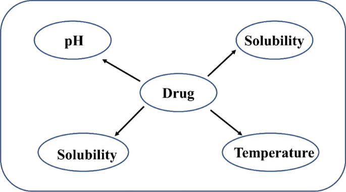
These are designed in a way to drug release within the definite period as a result of external stimulus [11, 13]. Figure 3 shows various triggered-based mechanisms employed for drug release in MSD.
pH-stimulated system
By altering the coating on the MDS, pH-dependent drug release is enabled.
Change of temperature
At room temperature, the entrapped drug may be too viscous to move rapidly from the microsponge to the dermis. However, when the temperature of the skin increases, the rate of active flow increases, and hence the rate of release increases as well.
Pressure
Applying pressure while rubbing can result in the release of drugs encapsulated in MDS onto the skin. The amount of gas released is governed by the sponge's quality. Microsponge formulations can be improved by altering the material used and a number of other variables.
Solubility
Water-miscible chemicals (antiperspirants and antiseptics) will release additives in the presence of water. Additionally, the diffusion mechanism may initiate the drug's release [14].
Engineering of MDS
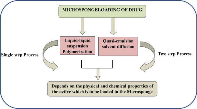
Apart from the microsponge composition, the delivery system's performance is influenced by the preparation processes used. However, due to their complexity and expense, microsponge preparation methods are restricted. Drug release can be performed in two ways: one-step process and two-step process (liquid–liquid polymerization) and quasi-emulsion diffusion method. Both methods are dependent on the physicochemical properties of the active drug that must be entrapped; for example, if the drug is a non-polar and inert material, it will produce a structure that is porosity, as shown in Fig. 4. Porogen drugs that do not inhibit polymerization and do not trigger it, as well as being stable towards free radicals, may be entrapped using a one-step procedure [3, 13].
Liquid–liquid suspension polymerization
Typically, a solution containing monomers and active additives that are water-immiscible is created first. Figure 5 summarises the number of stages involved in liquid–liquid suspension polymerization processes [15]. This water-soluble phase is suspended in water with agitation and often comprises chemicals such as emulsifiers and dispersants. Polymerization is achieved by activating monomers with the assistance of catalysis, temperature elevation, or irradiation after the solution has been formed with separate droplets of the desired size. The polymerization technique results in the formation of 1000 cages of MDS with spherical structures that are linked and resemble a bunch of grapes. After the polymerization is finished, the solid particles will be collected from the resulting suspension. After that, the particles were rinsed and dried before being used again [3, 16].
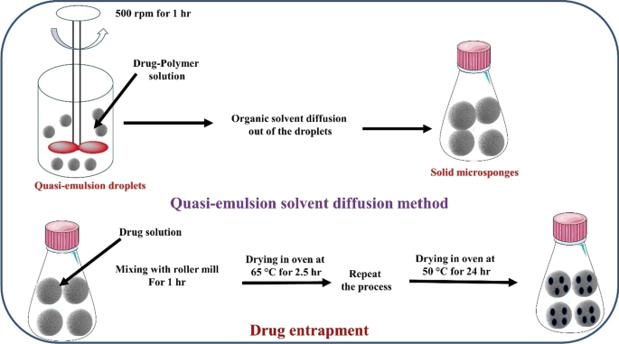
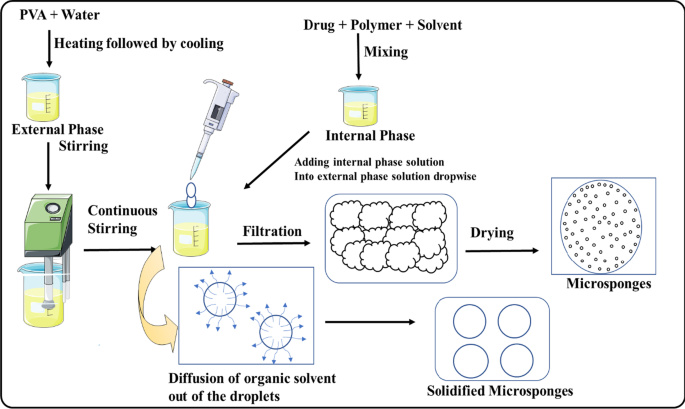
Quasi-emulsion solvent diffusion
The procedure described here is commonly used to make topical and oral microsponges. The procedure involves the production of two phases, one of which is the inner organic phase, which contains the drug, and the other is the outer aqueous phase, which is then agitated and filtered before being used. The inner phase is then blended drop by drop in the outer phase with the assistance of a mechanical stirrer for 60 min. The quasi-emulsion droplets were formed as a result of continual stirring, and the solid cages of microsponge were formed as a result of further organic solvent evaporation. After that, the microsponges were filtered and dried in the oven for 12 h. Figure 6 summarises the steps required in microsponge production utilising the approach quasi-emulsion solvent diffusion [17,18,19].
Multiple-emulsion solvent diffusion method
The approach was developed to create porous and biodegradable microspheres. An aqueous inner phase was used with the addition of stearyl amine, and the span was distributed in solution. This w/o emulsion is then dispersed again in an aqueous phase with polyvinyl alcohol to generate (w/o/w) double emulsion. This method reveals the advantage of capturing both soluble and insoluble actives. This method may also be used to entrap thermolabile compounds like proteins [3].
Addition of porogen
Porogens such as sodium bicarbonate or hydrogen peroxide were utilised to replace the numerous emulsions in this approach. To do this, the porogen was dissolved in a polymeric solution to create a single-phase system, which was then disseminated in a water phase containing polyvinyl alcohol. After that, an initiator was added to create numerous emulsions, and the organic solvent was evaporated, leaving the particles behind to build MDS [3].
Lyophilisation
The produced microspheres from the gelation procedure were converted into porous microspheres using this approach. After that, the microspheres were incubated in chitosan hydrochloride solution and stored for lyophilisation. This approach produces microsphere holes as a result of the quick solvent removal. Because of the quick elimination of solvent, the lyophilisation process of producing MDS has the disadvantage of creating shrunken or otherwise fragmented microparticles [3].
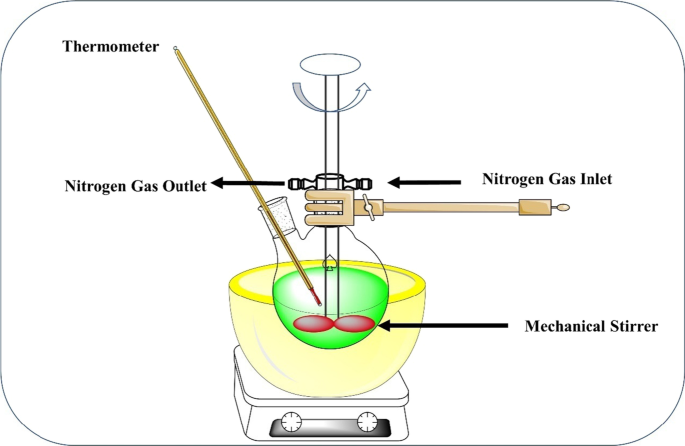
Ultrasound-assisted production
This approach was created by modifying the liquid–liquid suspension polymerization process for MDS generation. This approach uses beta-cyclodextrin monomer and diphenyl carbonate as a cross-linking agent to create MDS. To regulate the size of the microparticles, the reaction mixture is heated and sonicated. The mixture was cooled and powdered, which was then washed with distilled water before being washed with ethanol as illustrated in Fig. 7. This technique of production is also insufficient in terms of entrapping harmful cross-linking agent residue [3]. Table 1 compares the various strategies utilised in the production of MDS formulations.
Evaluation of MDS
Particle size determination
MDS should not have particles larger than 30 µm, with a typical particle size range of 10–25 µm. Laser light diffractometry or any equivalent technology is used to determine the particle size of loaded or unloaded MDS. Larger particles with a diameter of 30 µm cause grittiness; hence, tiny particles with a diameter of 10–25 µm are preferred for use in topical formulations [6, 8].
Surface morphology and topography
It can be used as a validation parameter to determine this since the produced MDS is coated with Au–Pd in an argon environment [8].
MDS loading efficiency
Equation 1 shows the loading efficiency of MDS [15]:
Equation 1 shows the production yield of MDS:
Determination of true density
The real density of MDS can be determined using an ultra-pycnometer under helium atmosphere [20].
Characterisation
The transport of drugs into the vehicle from MDS is also influenced by the pore diameter. The diameter (D) of the pore has an impact on the rate of drug release [5, 20]. Equation 3 shows the Washburn equation used to compute the pore diameter of MDS, where D is the pore diameter (μm); γ is the surface tension of mercury (485 dyn/cm); θ is the contact angle (130°); and P is the pressure (psia):
Porosity studies
To examine the size of nanocavities generated in MDS, a porosity analysis can be done. Pore structure, diameter, and volume can be calculated as per Eq. 4. Porosity can be determined as
Compatibility studies
TLC and FTIR both can be used to explore drug compatibility with reaction mixtures [8].
Resiliency
Resiliency of MDS also represents the viscoelastic nature and may be changed to make beadlets that are softer, as required [15].
In vitro dissolution studies
In vitro dissolution can be carried out with USP XXIII in dissolution assembly containing a 900 ml solution of saline phosphate buffer with a particular pH, heated to 375 °C, and spun at a set rotation rate per minute [21].
Stability studies
The investigations of stability are carried out in accordance with the International Council of Harmonization (ICH) criteria [21].
Polymer/monomer composition
The entrapped drug partition coefficient between the microsponge and the vehicle should be determined [6].
Studies on drug release and drug deposition
Typically, a Franz static diffusion cell is used to release the medicine from semi-solid dosage forms. In this procedure, the epidermal side of the skin is exposed to adequate temperature, while the dermal side, or skin inner side, is exposed to the receptor solution. The receptor compartment contains 20 mL of buffer media, which should be agitated at 600 rounds per minute and kept at 32 ± 0.5 °C. The skin was soaked with the diffusion medium for 1 h before the test sample was applied. A particular amount of the sample is put in the donor compartment. Diffusion cells are used to assess the amount of drug that is deposited on the skin, and they are subsequently deconstructed at specified time intervals. The skin is then gently removed, and the drug-affected area of the skin is cleansed with distilled water [5].
Formulation considerations
-
1.
MDS drug distribution system actions may then be used in a variety of goods such as lotions, creams, soaps, and powders. Certain thoughts are considered while developing the vehicle in order to attain the desired product features. The active substances' solubility in the vehicle should be kept to a minimum. If this is not the case, the vehicle will minimise the MDS activity [22].
-
2.
In order to minimise cosmetic issues, no more than 10–12% w/w MDS should be used in the car. Polymer designs and payloads for active MDS should be tuned for the needed release rate for the specified period. Some considerations should be made while developing vehicles for a specific application in order to attain the required product attributes.
-
3.
To avoid cosmetic issues, the powder load in the car should be sufficient (not more than 10–12%) [23]. The active agent's solubility in the vehicle should be limited; otherwise, the skin will degrade the polymer before it is delivered to it. For the release of the active agent throughout the specified duration, polymer design, as well as loading levels, must be suitably determined.
Safety considerations
Microsponge systems are built up of commonly used polymers with extremely low levels of remarkable monomers; also, cross-linking physiologically disables them, since the body is unable to biodegrade them and replicate them in other substances [24].
These microspheres are incredibly huge and pass through the stratum corneum due to their size. They linger under the skin's surface, gradually releasing their substance over time. This release pattern reduces the excessive build-up of active substances in the epidermis, perhaps improving topical drug safety [25].
Applications of MDS
Microsponge technology in cosmetics
Microsponge technology has an intriguing use in oral cosmetics, such as sustaining the release of volatile chemicals, therefore enhancing the duration of the ‘fresh feel’. Such volatile substances can be easily integrated into dental pastes or mouthwashes as MDS. MDS can be used in a range of coloured cosmetic items, such as rouge or lipsticks, to extend the life of the product by trapping the colour in the microsponge. MDS aids in the disintegration of uniformity and boosts the covering power. Hence, the colourful cosmetics made with MDS will be incredibly exquisite as a result [26].
MDS-based delivery systems for drug triggering
Because of the increased effectiveness of drugs when formulated as microsponge, improved safety, and better visual look of formulations used for topical treatments, over-the-counter drugs, and personal care items, microsponge delivery systems are being considered. This delivery method is designed to allow the active moiety to be released at a slower pace over time, perhaps reducing adverse effects while maintaining therapeutic efficacy. They can be used for a variety of purposes. It is mostly utilised for topical preparations, but it has also been employed recently for drug delivery via the oral route. It gives a formulator a variety of possibilities for making medicine or cosmetic product. They are made in such a way that they distribute active moiety efficiently with the least amount of dosage, as well as improve product stability, reduce adverse effects, and change drug release. Treatments including different moisturisers, sunscreens, and specific rejuvenate items are included in the microsponge delivery system that combines over-the-counter products [15, 27]. The microsponge system is used for topical administration in three ways in products that are in development or on the market:
-
1)
The active moieties are released for a prolonged period of time by the MDS reservoir.
-
2)
As a container for undesired things, such as excess skin oils, to be absorbed.
-
3)
MDS are closed containers that keep the components away from the skin while they work on the surface [15].
MDS are porous sponges that are used for topical treatment and, more recently, oral delivery of active ingredients. MDS is designed to administer a medicine efficiently with the smallest amount possible, as well as to increase product stability, reduce adverse effects, and change the release of a drug [28]. Table 2 summarises the available marketable items.
MDS for topical delivery
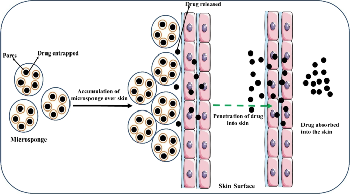
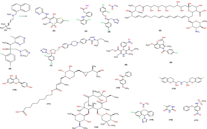
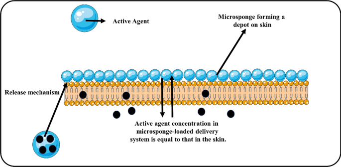
The action of traditional topical drug formulations is thought to be limited to the outermost layers of the skin. When these traditional items are put to the skin, they release their pharmaceutically active medicine, resulting in an extraordinarily concentrated film of pharmaceutically active drug that is rapidly absorbed. This excessive build-up of active components in the epidermis and dermis layers of the skin can be avoided by packaging the medicine in a microsponge drug delivery system (Fig. 8 and Table 3). This microsponge technology can significantly reduce drug irritation while maintaining its effectiveness. Furthermore, these drug-containing porous microspheres can be incorporated into dosage forms such as creams, gels, powders, and lotions [30, 31]
MDS for oral drug delivery
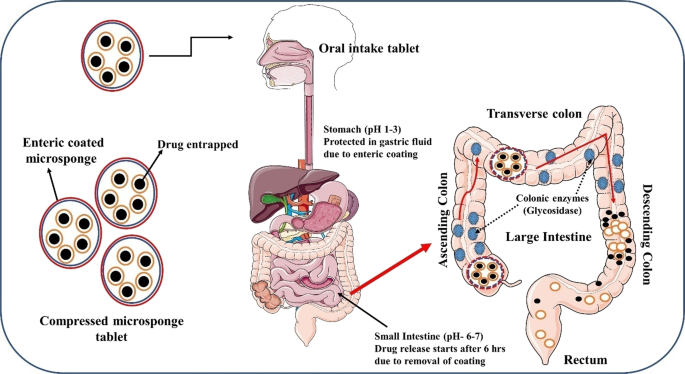
The oral administration of drug using MDS is appropriate because this technology has the ability to increase the release rate of drugs that are poorly soluble in water by entrapping these compounds in the pore system of the microsponge. The pH of the microsponge oral drug administration is regulated by it. Figure 9 shows that when the microsponge is removed from the mouth; the medicine is not delivered to the mouth and is protected by gastric juice (pH 1–3) owing to enteric coating. The coating dissolves in the gut owing to the action of colonic enzymes (glycosidases), and the medicine begins to distribute after 6 h. When MDS move through the descending colon of the big intestine, they transport the most drugs.
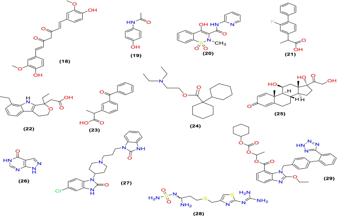
The microsponge delivery technology also has the potential to embrace drugs in a restricted environment and to transfer active ingredients to the lower region of the GIT in a regulated way. The fundamental reason for using the microsponge system as a carrier for colonic administration is that actives with a size of less than 200 µm can be easily taken up by macrophages in the colonic tissues, resulting in effective localised drug action at the targeted spot [19, 32]. Table 4 shows a list of drug candidates investigated using microsponge delivery for oral drug administration.
Ocular delivery
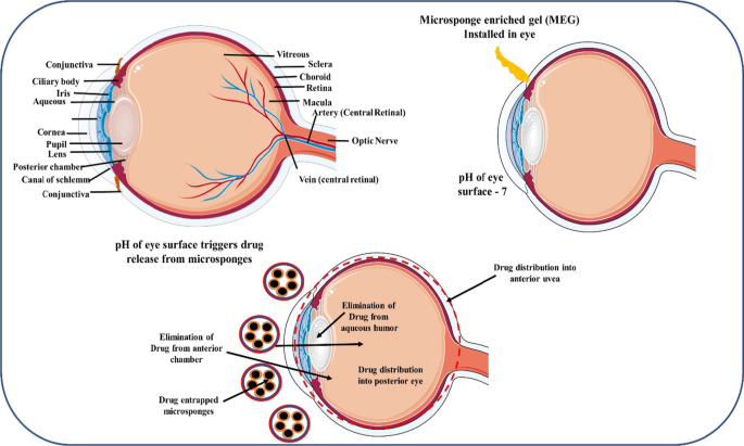
Water-soluble drugs can be applied topically as an ointment or an aqueous suspension, whereas water-insoluble pharmaceuticals can be applied topically as an ointment or an aqueous suspension. The pharmacokinetics of drug in the eye is a complicated process (Fig. 10). Following that, the drug is carried into the anterior chamber across the blood–aqueous barrier. The drug is transported from the anterior chamber to the Schlemm's canal and trabecular meshwork, where it is removed through aqueous humour turnover. Through the blood–aqueous barrier, the medicine is also removed from the aqueous humour and into the systemic circulation. Finally, a drug molecule in the blood crosses the blood–retina barrier and reaches the eye's posterior chamber [6].
MDS in cosmetics and dermatology
Won's initial patents for microsponge technology were issued to Advanced Polymer Systems, Inc. in 1987. Microsponge technology was used in cosmetics, over-the-counter (OTC), and prescription medicinal goods by this firm. MDS ensures drug localisation on the skin's surface and inside the epidermis, bridging the gap between systemic and local cutaneous side effects. Only the technological aspects of MDS for dermatological and cosmetic products differ. Cosmetic items have a lot shorter manufacture, marketing, and introduction period than dermatological products due to minimal regulatory restrictions. Table 5 and Fig. 11 list some examples of microsponge-based cosmetic products now available on the market [31].
Furthermore, skin targeting with microsponge drug delivery devices is possible, preventing excessive drug absorption into the percutaneous blood circulation. Table 6 and Fig. 12 show microsponge-based formulations for dermatological applications that have been produced using a variety of medicines [51].
MDS used for bone and tissue engineering
When polymethyl methacrylate powder was mixed with liquid methyl methacrylate monomer and tricalcium phosphate grains and calcium-deficient hydroxyapatite powders in water, the mixture formed bone-like compounds that looked like real bone. The final composites looked like they had holes in them and were like microsponges. The basic fibroblast growth factor (bFGF), which was incorporated into a collagen sponge sheet, was released into the mouse sub-cutis as the sponge matrix degraded. The bFGF had local angiogenic activity in a dose-dependent way. Microsponges of collagen with bFGF were injected into the hind limb of a mouse that had been deprived of blood. This caused a significant increase in blood flow, which could not have been achieved by giving the bFGF bolus. These findings show how important and useful type I collagen is as a source of bFGF [63, 64]. A biodegradable graft material with the collagen microsponge was made for cardiovascular tissue grafting because it would allow the tissue to grow back from scratch [65]. Using biodegradable polymer and collagen microsponge, an engineered patch is made that could be used to help repair the cardiovascular system. This patch could also be used as a new surgical material [65].
MDS in arthritis
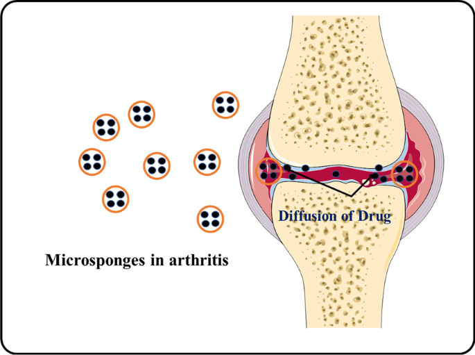
The use of a microsponge to dispense diclofenac has been studied in the treatment of arthritis (Fig. 13). Diclofenac is the most often prescribed nonsteroidal anti-inflammatory drug (NSAID) for the alleviation of pain and swelling associated with arthritis and other musculoskeletal disorders; however, it has been related to gastrointestinal irritation and first-pass metabolism. A topical treatment containing diclofenac MDS can be used to alleviate these concerns. Osmani et al. used a quasi-emulsion solvent diffusion technique to manufacture MDS gels containing diclofenac diethylamine to achieve a prolonged release for arthritis therapy. They compared their findings to the commercial Voltaren Emulgel 1.16 per cent w/w. The gel only released 81.11 per cent of the medicine in 4 h, but the microsponge-based gel released the drug over 8 h [62]. Hadi et al. produced MDS containing lornoxicam as the active component for treating arthritis and integrated them into tablet form, finding that the drug was released for a long duration, ranging from 86% to 96 per cent to 12 h [66].
Skin protection using MDS
UV radiation may cause sunburns and malignancies like basal carcinoma and malignant melanoma; hence, sunscreens are used to protect against it. To improve the impact, MDS can be employed. MDS was developed by Pawar et al. to deliver oxybenzone, a broad-spectrum sunscreen chemical, to the skin. To make it, they employed a quasi-emulsion solvent diffusion method. When applied as a lotion or cream, oxybenzone produces skin irritation, dermatitis, and systemic absorption. They employed 32 factorial designs to maximise the influence of ethyl cellulose and dichloromethane and then dispersed it in hydrogel for future research. The entrapment efficiency of the revised formulation was determined to be 96.9% 0.52 per cent. The gel released in a controlled manner and did not irritate the rats' skin. The highest recovery was seen during the creep recovery test, suggesting significant elasticity. In comparison with the one on the market, the formulation had a better sun protection factor, as well as less irritation and toxicity. The microsponge gel had an SPF of 25, but the marketed lotion had an SPF of 20, which might be due to the microsponge gel's slower drug release, which resulted in prolonged oxybenzone retention [45].
MDS in acne
Acne is a common skin problem that is frequently accompanied by skin inflammation. Benzoyl peroxide is the most common drug used to treat it. Jelvehgari and his colleagues used ethyl cellulose MDS on the skin to inhibit the release of benzoyl peroxide. Drug release was greater for the first hour and then stayed consistent for the next 7 hours, according to research. This might be owing to the existence of a nonencapsulated drug, which once released became constant, suggesting that the entrapped drug had been freed [67]. Osmani et al. produced a cream using miconazole nitrate MDS, an anti-acne agent, to achieve prolonged release. The Eudragit RS-100 MDS were created using a quasi-emulsion solvent diffusion method. MDS was made and incorporated into a cream. Drug-loaded MDS released 78.28% of the drug for up to 8 h, whereas traditional creams were exhausted after 4 h, releasing only 83.09 per cent of the drug [62].
MDS in psoriasis
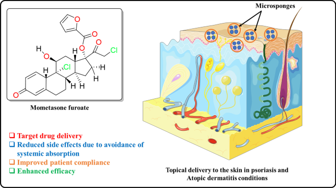
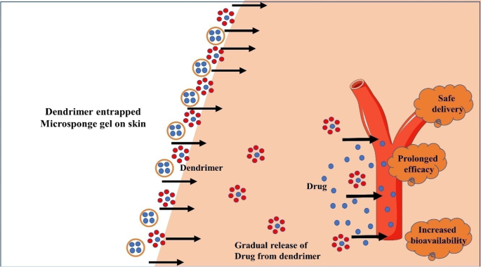
Psoriasis is a chronic inflammatory disorder that affects the skin. It has a negative impact on the quality of life of sick person. MDS has also been studied as a treatment for psoriasis. To make MDS for the drug mometasone furoate, an emulsion solvent diffusion method is employed (Fig. 14). Mometasone furoate is used to treat inflammatory and pruritic diseases like psoriasis. MDS was created using the emulsion solvent diffusion process. The release profiles demonstrated a biphasic release with an initial burst effect. 29–36% of the medicine was released in the first hour, and 78–95% of the drug was released after 8 h [44]. Due to the continuous release of drug, a microsponge gel containing clobetasol propionate was used for psoriasis therapy to reduce the number of applications necessary. They discovered that drug release might last up to 12 h, versus 2.5 h in the usual form [44]. Dithranol microsponge gel is encapsulated in a dendrimer for efficient topical therapy and useful in psoriasis (Fig. 15) [68].
MDS in skin infections
MDS is also used to treat eczema and atopic dermatitis in the form of gels or lotions. Mupirocin is a topical antibiotic that is used to treat both primary and secondary skin infections. Mupirocin MDS was developed by Amrutiya et al. for long-term usage against skin infections. They used an emulsion solvent diffusion approach to generate mupirocin MDS, which they then put into an emulgel foundation. Researchers were able to demonstrate controlled drug release using a cellulose dialysis membrane. Figure 16 shows that a considerable proportion of the medicine persisted in the skin for up to 24 h after release and drug deposition investigations on the rat belly skin. The increased formulations were shown to be stable and non-irritating in a Draize patch test. The drug release from the ointment lasted up to 4 h, whereas the release from the microsponge gel lasted up to 10 h [44].
Diabetic wound healing using MDS
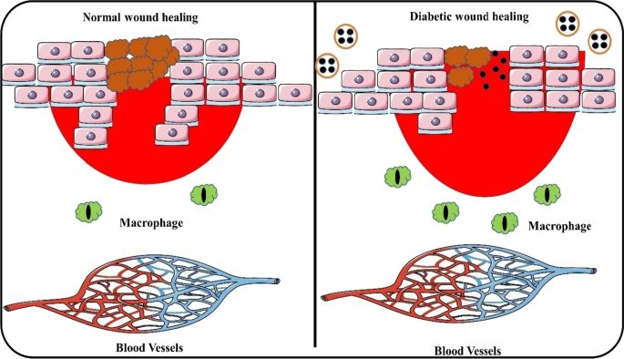
Pandit et al. made nebivolol-loaded MDS and encapsulated them in gel to keep the wound moist during the last stages of healing. Taking nebivolol, which is an antihypertensive drug, dilates the blood vessels. In diabetic wounds, the nitric oxide route reduces diabetic neuropathy and improves the function of the endothelial cells in the wound. It was confirmed from in vitro test that after 8 hours, about 80% of the drug was released. Due to the microsponge gel, the release of the drug has been slowed down. Because of the porous nature of the formulation, diabetic rats show quick wound healing (Fig. 17) [46].
MDS as antifungal
To make MDS of terbinafine hydrochloride, an antifungal drug, Thavva et al. employed a quasi-emulsion solvent diffusion technique. In vitro, the drug-loaded microsponge-based gel was compared to a drug-loaded plain gel. The drug-loaded plain gel released 96.65 per cent of the drug for up to 6 h, but the microsponge-loaded gel released the drug optimally for up to 10 h. Terbinafine hydrochloride-loaded microsponge gels had a longer drug release than conventional gels [33]. Dombe et al. created oxiconazole nitrate MDS using a quasi-emulsion solvent diffusion method, which they then incorporated into the gel. The antifungal oxiconazole nitrate has limited aqueous solubility, as well as side effects and unpleasant responses. The MDS produced were distinct and spherical in shape, with a high production yield of 61.44–80.45%, showing that this method of preparation was a good strategy for microsponge manufacturing. The highest drug release was found to be 87.77 per cent during an 8-h period. On a microsponge gel, the drug was released in a regulated manner for 12 h [37, 51].
MDS in melanoma
Grimes PE created hydroquinone MDS with 4 per cent hydroquinone and 0.15 per cent retinol for the treatment of melanoma and post-inflammatory hyperpigmentation (PIH). MDS that releases hydroquinone was created to give long-term drug delivery with little skin irritation. It was decided to conduct an open-label experiment. Improvements in sickness symptoms and pigmentation intensity were statistically significant as compared to baseline at weeks 4, 8, and 12 (p < 0.001). The lesion area and colorimetry measurements both improved significantly with each visit (p < 0.001). The formulation was well tolerated, with only one patient dropping out owing to a non-life-threatening allergic reaction [69].
MDS in colon cancer
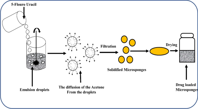
MDS can be used to decrease the toxicity and long-term release of oral drugs. Gupta and his team came up with Eudragit RS100-based 5-Fluorouracil MDS to treat colon cancer. There are a lot of different types of solid tumours that 5-FU can be used to treat. 5-FU can be more effective if it has a higher relative concentration in tumours. As a result, the toxicity level goes down. Oil-in-oil emulsion solvent diffusion was used to make MDS. Pure 5-FU was found to be released in about 20 min, but the MDS makes the release time last for about 5 h. It has been found that when it comes to the viability of cells, MDS-loaded 5-FU is better than 5-FU on its own. In the study, it was found that 5-FU MDS can be used instead of oral 5-FU to treat cancer (Fig. 18) [70].
Other applications
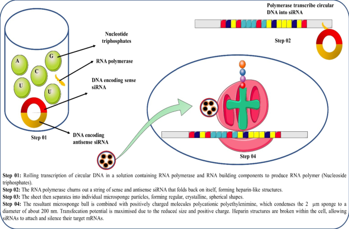
MDS has emerged as a novel drug delivery technology that may be utilised to deliver siRNA and fibroblast growth factor in addition to topical and oral administration. Because a single RNAi microsponge may transfer more than half a million copies of siRNA to a cell, this method of siRNA administration can be used and extended in the field of modern therapeutics and pharmacological research. Researchers employed a new approach that combines femtosecond X-ray single-shot diffraction with an X-ray free-electron laser and coherent diffraction imaging with synchrotron X-rays to explore the nanostructure formation involved in RNA interference microsponge. Figure 19 depicts the mechanistic transfer of siRNA from a microsponge [6, 71].
Recent advances in the MDS
Advanced formulations are being developed by several researchers and pharmaceutical businesses. They are as follows, with a higher level of stability than MDS.
-
1)
Nanosponges These have been shown to be excellent carriers for gas transport. When cytotoxic is combined with nanosponges as carrier systems, the efficacy of the drug is increased, by allowing it to be utilised to target cancer cells. Cross-linking the β-CD molecule with biphenyl carbonate produces β-CD nanosponges. They can be utilised to make both hydrophilic and hydrophobic medicines. Flurbiprofen, dexamethasone, itraconazole, and other drugs were examined using this sophisticated approach [72].
-
2)
Nanoferrosponges Nanoferrosponges are self-promising carriers with increased penetration towards a particular location due to an external magnetic reaction that allows carriers to penetrate deeper tissue before magnetic material is excluded, leaving a porous system behind.
-
3)
Porous microbeads Porous microbeads were created as a result of better microsphere properties. Polymerization and cross-linking processes are utilised to create solid porous microbeads. The monomer used in the high internal phase emulsion approach has an exterior oil phase, an internal aqueous phase, and a cross-linker. Topical, oral, and buccal drug delivery systems all use microbeads. Due to increased RNA stability and effective siRNA encapsulation, this technique provides a novel therapeutic pathway for siRNA administration [73].
Future perspectives of microsponge drug delivery
Because of its diverse uses in pharmaceuticals and its peculiar porosity system properties, the microsponge drug delivery system will have a very bright future in the next years. One of the primary problems for this drug delivery system in the future will be the oral delivery of peptides, which can be accomplished by using core delivery of drug-loaded MDS and varying and applying the number of polymer ratios. As a result, the microsponge drug delivery system is a very promising subject in the future that should be investigated for its potential benefits [17].
In the near future, MDS devices might be investigated for tissue engineering. Tissue engineering has a lot of promise for improving patients' quality of life. A good example is vascular tissue engineering. The traditional strategy is to employ an extended type of polytetrafluoroethylene, which is used therapeutically to recreate bigger diameter arteries; however, this method is not suitable for tiny arteries since they are seen as foreign material by the body, causing thrombosis. As a result, a bypass mechanism is necessary. A unique intended vascular graft with tiny calibre made of polyglycolic acid and polylactic acid was constructed utilising a scaffold design process and provided with microsponge to shape a vascular patch, which was subsequently tested on dogs. And the results revealed that the dogs healed completely without any complications and that there was no need to employ an anti-thrombotic drug with the graft because it is comprised of the biodegradable polymer [5, 6]. The true difficulty of drug delivery via microsponge system in the near future is achieving core/shell delivery of active loaded MDS for oral administration of peptides by changing the polymer ratio [73].
MDS is a novel drug delivery system with unique properties such as enhanced product performance and elegancy, extended release, and improved drug release profile, reduced irritation, improved physical, chemical, and thermal stability, which makes it flexible to develop novel product forms. Biodegradable polymers can also be employed for tissue engineering and regulated oral drug administration. It offers a wide range of benefits when it comes to formulation. Alternative drug delivery methods, such as parenteral and pulmonary, must also be developed for MDS. These carrier systems were used in cosmetics because of their elegance. These advancements allowed researchers to use them in a variety of ways. These formulation innovations also open up new avenues for drug delivery. As a result, microsponge-based drug delivery technology is expected to become a viable drug delivery matrix material in the future for a variety of therapeutic applications [74, 75].
Conclusions
Microsponge technology and the adaptability it provides have a lot of promise in the market because of the desire for novel and highly efficient pharmaceutical and cosmetic goods. MDS is a promising technology for the highly regulated release of an active chemical loaded in MDS, which also leads to a decrease in pharmacological side effects while maintaining therapeutic efficacy. It also exhibited considerable improvements in formulation stability, as well as increased formulation elegance and flexibility. Based on a number of studies, they are also thought to be non-toxic, non-allergic, and non-mutagenic in nature. This drug delivery mechanism is now employed mostly in over-the-counter (OTC) skincare products, prescription medicines, cosmetics, and sunscreens. As a result of its diverse possibilities in drug administration, it is a very promising technology that is likely to be thoroughly examined in the next years through a variety of research projects.
Availability of data and materials
Data and material will be available on request.
Abbreviations
- 5-FU:
-
5-Flouro uracil
- MDS:
-
Microsponges delivery system
- OTC:
-
Over-the-counter
- RNA:
-
Ribonucleic acid
- API:
-
Active pharmaceutical ingredient
- XRD:
-
X-ray diffraction method
- DSC:
-
Differential scanning calorimetry
- PIH:
-
Post-inflammatory hyperpigmentation
References
Naga Jyothi K, Dinesh Kumar P, Arshad P, Karthik M, Panneerselvam T (2019) Microsponges: a promising novel drug delivery system. J Drug Deliv Ther 9(5-s):188–194. https://doi.org/10.22270/jddt.v9i5-s.3649
Singhvi G, Manchanda P, Hans N, Dubey SK, Gupta G (2018) Microsponge: an emerging drug delivery strategy. Drug Dev Res 80(2):200–208. https://doi.org/10.1002/ddr.21492
Sharma S, Sharma A, Kaur C (2020) MDS: as a topical drug delivery system. Int J Pharm Sci Res 11(2):524–534
Mohite PB, Khanage SG, Harishchandre VS, Shirshath Y (2016) Recent advances in microsponge drug delivery system. J Crit Rev 3(1):9–16
Ahmed A, Makram M, Sayed M, Louis D (2018) An overview of microsponge as a novel tool in drug delivery. MADD 2(3):1–7. https://doi.org/10.31031/MADD.2018.02.000537
Kumari A, Jain A, Hurkat P, Tiwari A, Jain SK (2018) Eudragit S100 coated MDS for Colon targeting of prednisolone. Drug Dev Ind Pharm 44(6):902–913. https://doi.org/10.1080/03639045.2017.1420079
Mandava SS, Thavva V (2012) Novel approach: microsponge drug delivery system. Int J Pharm Sci Res 3(4):967–980. https://doi.org/10.13040/IJPSR.0975-8232.3(4).967-80
Vitthal P, Anuradha S (2020) A review on MDS drug delivery system. Int J Res Anal Rev 7(1):961–974
Ghadge M, Purakasythya D, Pramanik A, Garg SK (2018) Microsponge – aeon in the field of topical formulation. Int J Pharm Pharm Sci 14(1):39–54
Jagtap SC, Karale AA, Ambekar AW (2014) Microsponge: a novel topical drug delivery system. J Drug Deliv 3(4):1–9
Rajeshwari S, Swapna V (2019) MDS as a neoteric cornucopia for drug delivery systems. Int J Curr Pharm Res 11(3):4–12
Kumar S, Tyagi LK, Singh D (2011) Microsponge delivery system (MSD): a unique technology for delivery of active ingredients. Int J Pharm Sci Res 2(12):3069–3080. https://doi.org/10.13040/IJPSR.0975-8232.2(12).3069-80
Ravi V, Ravi G, Bose PSC, Saritha D (2019) MDS- a comprehensive review: success and challenges. Indo Am J Pharm 9(7):3056–3067
Tiwari A, Mishra MK, Shukla A, Yadav SK (2016) Microsponge: an augmented drug delivery system. Am J Pharm Tech Res 6(6):79–95
Kar AK, Kar B, Parya H, Kundu S, Hirawat R (2018) A novel approach on microsponge: multifunctional modern dosage form. Int J Pharm Sci Rev Res 51(2):64–72
Kapoor D, Vyas RB, Lad C, Patel M, Tyagi BL (2014) A review on microsponge drug delivery system. J Drug Deliv Ther 4(5):29–35
Borawake PD, Kauslya A, Shinde JV, Chavan R (2021) Microsponge as an emerging technique in novel drug delivery system. J Drug Deliv Ther 11(1):171–181. https://doi.org/10.22270/jddt.v11i1.4492
Chadawar V, Shaji J (2007) Microsponge delivery system. Curr Drug Deliv 4(2):123–129
Srivastava R, Pathak K (2015) Microsponges: a futuristic approach for oral drug delivery. Expert Opin Drug Deliv 9(7):863–878. https://doi.org/10.1517/17425247.2012.693072
Balamurugan K, Kshirasagar GP (2019) Microsponge: as a drug delivery system. J Pharm Innov 8(1):139–143
Sumbria R, Gupta NB, Bhandari N, Kumar A, Mahajan S (2017) Microsponges: topical preparation and its applications. World J Pharm Pharm Sci 6(4):629–642
Chandramouli Y, Shaik Firoz B, Yasmeen R, Vikram A, Mahitha AU (2012) Microsponges: a novel drug delivery system for controlled delivery of topical drugs. Int J Pharm Res Appl 2(2):79–86
Kircik LH (2013) The microsponge delivery system reduces facial oiliness and shine during acne therapy. J Drugs Dermatol 12(11):1268–1270
Pandit AP, Patel SA, Bhanushali VP, Kulkarni VS, Kakad VD (2016) Nebivolol-loaded microsponge gel for healing of diabetic wound. AAPS Pharm SciTech 18(3):846–854. https://doi.org/10.1208/s12249-016-0574-3
Mantry S, Bagchi A, Das S (2015) Microsponge as a novel strategy of drug delivery system. Univers J Pharm Res 1(1):32–38
Vishwakarma P, Choudhary R (2019) MDS: a novel strategy to control the delivery rate of active agents with reduced skin irritancy. J Drug Deliv Ther 9(6-s):238–247
Tile MK, Pawar AY (2015) MDS: a novel strategy for drug delivery. Int J Pure Appl Biosci 3(1):224–235
Pradhan SK (2011) MDS as the versatile tool for drug delivery system. Int J Res Pharm 1(2):243–258
Kaity S, Maiti S, Ghosh AK, Pal DK, Ghosh A, Banerjee S (2010) MDS: a novel strategy for drug delivery system. J Adv Pharm Technol 1(3):284–290. https://doi.org/10.4103/0110-5558.72416
Shaha V, Jain H, Krishna J, Patel P (2010) Microsponge drug delivery: a review. Int J Res Pharm Sci 1(2):212–218
Mahant S, Kumar S, Nanda S, Rao R (2020) MDS for dermatological applications: perspectives and challenges. Asian J Pharm Sci 15(3):273–291. https://doi.org/10.1016/j.ajps.2019.05.004
Shrivastava S, Kumar D, Dubey CK, Singh SP, Khinchi MP (2017) A review: microsponge- an effective drug delivery system. Asian J Pharm Res Dev 5(2):1–8
Thavva V, Baratam SR (2019) Formulation and evaluation of terbinafine hydrochloride microsponge gel. Int J Appl Pharm 11(6):78–85. https://doi.org/10.22159/ijap.2019v11i6.32502
He Y, Majid K, Maqbool M, Hussain T, Yousaf AM et al (2020) Formulation and characterization of lornoxicam-loaded cellulosic-microsponge gel for possible applications in arthritis. Saudi Pharm J 28(8):994–1003. https://doi.org/10.1016/j.jsps.2020.06.021
Thiresssa B, Prasad AR, Haroled PPL (2018) Formulation and evaluation of lornoxicam MDS using eudragit RS 100 and eudragit RSPO. Asian J Pharm Clin Res 11(10):217–221
Maiti S, Kaity S, Ray S, Sa B (2011) Development and evaluation of xanthan gum-facilitated ethyl cellulose MDS for controlled percutaneous delivery of diclofenac sodium. Acta Pharm 61(3):257–270. https://doi.org/10.2478/v10007-011-0022-6
Yadav V, Jadhav P, Dombe S, Bodhe A, Salunkhe P (2017) Formulation and evaluation of microsponge gel for topical delivery of antifungal drug. Int J Appl Pharm 9(4):30. https://doi.org/10.22159/ijap.2017v9i4.17760
Bansode AS, Kute BV, Vethekar VS, Kote SP, Varhadi MK et al (2019) Formulation development and evaluation of microsponge loaded topical gel of nystatin. J Drug Deliv Ther 9(2-s):451–461. https://doi.org/10.22270/jddt.v9i2-s.2567
Mohan DS, Gupta VRM (2019) Microsponge based drug delivery system of voriconazole for fungal infection: formulation development and In-vitro evaluation. J Drug Deliv Ther 9(3):369–378. https://doi.org/10.22270/jddt.v9i3.2840
Monika DJS, Prasad DN, Hans M, Kumari S (2019) Preparation and characterization of itraconazole MDS using eudragit RSPO and study the effect of stirring on the formation of microsponges. J Drug Deliv Ther 9(3-s):451–458. https://doi.org/10.22270/jddt.v9i3-s.2879
Yang M, Cui F, You B, Fan Y, Wang L, Yue P, Yang H (2003) Preparation of sustained-release nitrendipine microspheres with Eudragit RS and Aerosil using quasi-emulsion solvent diffusion method. Int J Pharm 259(1–2):103–113. https://doi.org/10.1016/s0378-5173(03)00209-6
Mahajan AG, Jagtap LS, Chaudhari AL, Swami SP, Mali PR (2011) Formulation and evaluation of microsponge drug delivery system using indomethacin. Int Res J Pharm 2(10):64–69
Nagula RL, Wairkar S (2020) Cellulose MDS based gel of naringenin for atopic dermatitis: design, optimization, in vitro and in vivo investigation. Int J Biol Macromol 164:717–725. https://doi.org/10.1016/j.ijbiomac.2020.07.168
Amrutiya N, Bajaj A, Madan M (2009) Development of MDS for topical delivery of mupirocin. AAPS Pharm SciTech 10(2):402–409. https://doi.org/10.1208/s12249-009-9220-7
Pawar V, Salunkhe A (2020) A review on MDS drug delivery system. Int J Res Anal Rev 7(1):961–974
Pandit AP, Patel SA, Bhanushali VP, Kulkarni VS, Kakad VD (2017) Nebivolol-loaded microsponge gel for healing of diabetic wound. AAPS Pharm SciTech 18(3):846–854. https://doi.org/10.1208/s12249-016-0574-3
Ravi R, Kumar SKS, Parthiban S (2013) Formulation and evaluation of the MDS gel for an anti-acne agent for the treatment of acne. Int J Pharm Sci Res 3(1):32–38
Bothiraja C, Gholap AD, Shaikh KS, Pawar AP (2014) Investigation of ethyl cellulose microsponge gel for topical delivery of eberconazole nitrate for fungal therapy. Ther Deliv 5(7):781–794. https://doi.org/10.4155/tde.14.43
Jain SK, Kaur M, Kalyani P, Mehra A, Kaur N, Panchal N (2019) MDS enriched gel for enhanced topical delivery of 5-fluorouracil. J Microencapsul 36(7):677–691. https://doi.org/10.1080/02652048.2019.1667447
Singh RP, Singh S, Prajapati SK, Saxena GK (2013) Nimesulide loaded MDS: in-vitro and ex-vivo evaluation. Int J Biopharm 4(1):49–60
Barde PM, Basarkar GD (2015) Formulation, development and in vitro evaluation of terbinafine HCl microsponge gel. Int J Pharm Sci Rev Res 32:310–314
Bhatia M, Saini M (2018) Formulation and evaluation of curcumin microsponge for oral and topical drug delivery. Prog Biomater 7:239–248. https://doi.org/10.1007/s40204-018-0099-9
Jain V, Singh R (2011) Design and characterization of colon-specific drug delivery system containing paracetamol microsponge. Arch Pharm Res 34(5):733–740. https://doi.org/10.1007/s12272-011-0506-4
Rajab NA, Jawad MS (2016) Formulation and in vitro evaluation of piroxicam microsponge as a tablet. Int J Pharm Pharm Sci 8(2):104–114
Orlu M, Cevher E, Araman A (2006) Design and evaluation of colon specific drug delivery system containing flurbiprofen MDS. Int J Pharm 318(1–2):103–117. https://doi.org/10.1016/j.ijpharm.2006.03.025
Swetha A, Gopal RM, Venkata RK, Niyaz Basha B, Kotireddy V (2011) Formulation and in vitro evaluation of etodolac entrapped in microsponge based drug delivery system. Int J Pharm 1(2):73–80
Comoglu T, Savaser A, Ozkan Y, Gonul N, Baykara T (2007) Enhancement of ketoprofen bioavailability by formation of microsponge tablets. Pharmazie 62:51–54. https://doi.org/10.1691/ph.2007.1.6016
Jain V, Singh R (2010) Dicyclomine-loaded eudragit®-based microsponge with potential for colonic delivery: preparation and characterization. Trop J Pharm Res. https://doi.org/10.4314/tjpr.v9i1.52039
Sonali SRP, Prajapati SK (2014) Formulation and evaluation of prednisolone loaded MDS for colon drug delivery: in-vitro and pharmacokinetic study. Int J Pharm Sci Res 5(5):1994–2005. https://doi.org/10.13040/IJPSR.0975-8232.5(5).1994-05
Patel D, Gohil D, Patel D, Sheth H, Patel S, Pandya K, Shah C (2016) Formulation and evaluation of floating MDS of allopurinol. Int J Pharm Sci Res 7(3):135–154
Charagonda S, Puligilla R, Ananthula M, Bakshi V (2016) Formulation and evaluation of famotidine floating MDS. Int Res J Pharm 7(4):62–67. https://doi.org/10.7897/2230-8407.07440
Osmani R, Aloorkar N, Thaware B, Kulkarni P, Moin A, Hani U (2015) Microsponge based drug delivery system for augmented gastroparesis therapy: formulation development and evaluation. Asian J Pharm Sci 10:442–445. https://doi.org/10.1016/j.ajps.2015.06.003
Chen G, Sato T, Ohgushi H, Ushida T, Tateishi T, Tanaka J (2005) Culturing of skin fibroblasts in a thin PLGA-collagen hybrid mesh. Biomaterials 26:2559–2566
Kanematsu A, Marui A, Yamamoto S, Ozeki M, Hirano Y, Yamamoto M et al (2004) Type I collagen can function as a reservoir of basic fibroblast growth factor. J Control Release 94:281–292
Iwai S, Sawa Y, Ichikawa H, Taketani S, Uchimura E, Chen G et al (2004) Biodegradable polymer with collagen microsponge serves as a new bioengineered cardiovascular prosthesis. J Thorac Cardiovasc Surg 128:472–479
Hadi MA, Rao NG, Rao AS (2015) Formulation and evaluation of mini-tablets-filled-pulsincap delivery of lornoxicam in the chronotherapeutic treatment of rheumatoid arthritis. Pak J Pharm Sci 28(1):185–193
Jelvehgari M, Siahi-Shadbad MR, Azarmi S, Martin GP, Nokhodchi A (2006) The microsponge delivery system of benzoyl peroxide: preparation, characterization and release studies. Int J Pharm 308(1–2):124–132. https://doi.org/10.1016/j.ijpharm.2005.11.001
Tripathi PK, Gorain B, Choudhury H, Srivastava A, Kesharwani P (2019) Dendrimer entrapped microsponge gel of dithranol for effective topical treatment. Heliyon 5:e01343. https://doi.org/10.1016/j.heliyon.2019.e01343
Grimes PE (2004) A microsponge formulation of hydroquinone 4% and retinol 0.15% in the treatment of melasma and post-inflammatory hyperpigmentation. Cutis 74:362–368
Gupta A, Tiwari G, Tiwari R, Srivastava R (2015) Factorial designed 5-fluorouracil-loaded MDS and calcium pectinate beads plugged in hydroxypropyl methylcellulose capsules for colorectal cancer. Int J Pharm Investig 5(4):234–246
Han S, Kim H, Lee JB (2017) Library siRNA-generating RNA nanosponges for gene silencing by complementary rolling circle transcription. Sci Rep 7(1):1–7. https://doi.org/10.1038/s41598-017-10219-y
Garud ST, Tiwari K (2018) Microsponges: a novel approach. Asian J Pharm Sci Tech 8(1):1–9
Valluru R, Ravi G, Bose SP, Damineni S (2019) Microsponges- a comprehensive review: success and challenges. Indo Am J Pharm Res 9(7):3056–3067
Verma R, Kaushik D (2020) Design and optimization of candesartan loaded self-nanoemulsifying drug delivery system for improving its dissolution rate and pharmacodynamic potential. Drug Deliv 27(1):756–771. https://doi.org/10.1080/10717544.2020.1760961
Verma R, Kaushik A, Almeer R, Rahman MH, Abdel-Daim MM, Kaushik D (2021) Improved pharmacodynamic potential of rosuvastatin by self-nanoemulsifying drug delivery system: an in vitro and in vivo evaluation. Int J Nanomedicine 16:905–924. https://doi.org/10.2147/IJN.S287665
Acknowledgements
The authors acknowledge the Dean, Faculty of Pharmacy, IFTM University, Moradabad, for providing continuous support for the article.
Funding
There was no particular grant for this assessment from any funding source in the public, commercial, or non-profit sectors.
Author information
Authors and Affiliations
Contributions
VT and AT contributed to conceptualization; BP, VT and AT helped in writing—original draft preparation; AT and MK performed writing—review and editing. All authors have read and approved the final manuscript.
Corresponding author
Ethics declarations
Ethics approval and consent to participate
Not applicable.
Consent for publication
Not applicable.
Competing interests
The authors declare that they have no conflict of interest.
Additional information
Publisher's Note
Springer Nature remains neutral with regard to jurisdictional claims in published maps and institutional affiliations.
Rights and permissions
Open Access This article is licensed under a Creative Commons Attribution 4.0 International License, which permits use, sharing, adaptation, distribution and reproduction in any medium or format, as long as you give appropriate credit to the original author(s) and the source, provide a link to the Creative Commons licence, and indicate if changes were made. The images or other third party material in this article are included in the article's Creative Commons licence, unless indicated otherwise in a credit line to the material. If material is not included in the article's Creative Commons licence and your intended use is not permitted by statutory regulation or exceeds the permitted use, you will need to obtain permission directly from the copyright holder. To view a copy of this licence, visit http://creativecommons.org/licenses/by/4.0/.
About this article
Cite this article
Tiwari, A., Tiwari, V., Palaria, B. et al. Microsponges: a breakthrough tool in pharmaceutical research. Futur J Pharm Sci 8, 31 (2022). https://doi.org/10.1186/s43094-022-00421-9
Received:
Accepted:
Published:
DOI: https://doi.org/10.1186/s43094-022-00421-9
